
What is cone beam computed tomography?
An X ray that allows 3 dimensional images of the head.
Why is it important?
Visualization in three dimensions at Strathcona dental has provided 3 important functions:
- Allows close to pinpoint accuracy in the placement of dental implants.
- Evaluate vital structures prior to surgery.
- Screening for harmful pathology not easily picked up by traditional 2d imaging.
Should I be concerned about the Radiation dose?
The iCAT can be calibrated to different radiation doses for different applications. We use the low-resolution setting, it is equivalent to the 2d panoramic images of the past. The dosage is minimal.
Do all offices have CBCT imaging?
It has become much more common in dental offices today compared to 5 years ago. Especially if surgery and dental implants are being performed. At Strathcona dental we provide this high value imaging at the same price as 2d images. Easily 25% less than a private radiation facility. We perform CBCT imaging every 5 years instead of panoramic images.
The images below are examples of pathology that have been found over the last year, usually on routine exams. Without Cone beam CT most of the pathology would go unnoticed. Patient names have been removed for privacy. Thank you for allowing the use to educate our patient base on the benefits of this imaging modality.
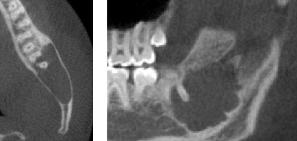
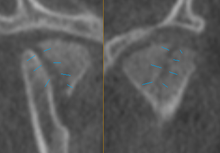
Case 1: Traumatic bone cyst that was successfully removed by oral surgeon.
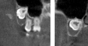
Case 2: Dentigerous cyst around impacted wisdom tooth. Referred to surgeon.
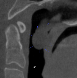
Case 3: Patient concern with breathing issues. A large mass was discovered behind the soft palate. It could not be visualized in the mouth. It was surgically removed and biopsied as benign with no further concerns.
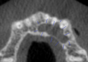
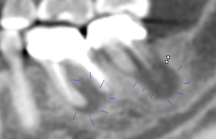
Case 4: New patient exam uncovered a nasopalatine duct cyst behind the front top teeth going to the premolars. Oral surgeon referral for removal.
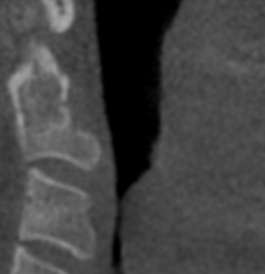
Case 5: New patient (5 yrs old) fell off bike. Did not complain of unusual discomfort. Imaging found significant condylar (jaw) fractures on both sides of the jaw. Referral to oral surgeon was made.
Case 6: Significant narrowing of lower airway noticed. Biopsy identified cancer. Surgery and treatment commenced.
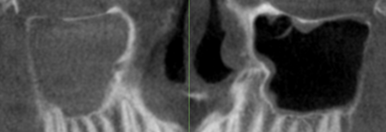
Case 7: It is common to see sinusitis. When the sinus is chronically full with inflamed tissue it leads to pressure pain, blocked passages and higher incidence of infection. Clear sinus on the right is black. Fully occluded sinus on left needs ears nose throat specialist referral.
Case 8: CBCT is used weekly to evaluate root end abscess of dead, infected and broken teeth.
It would be hard to imagine practicing dentistry without this useful tool. We have had one in office for over 10 years. We see the value in taking them routinely every 5-10 years.
Best regards